Family owned and trusted since 1983

S2K Commerce - Products Dropdown
Actions
S2K Commerce - Shopping Cart
Actions
S2K Commerce - Order Entry
Actions
Laboratory Equipment and Scientific Research Supplies
Search our wide selection of products: Made in the USA, Cell and Tissue Culture, PCR, Liquid Handling, Government, and more.


Stocked products ship in 24 hours

Responses within 24 hours
Featured Categories
-

Cell / Tissue Culture
TPP Plates, flasks, dishes, bioreactors, filtration, & more. Flatter surfaces, better treatments, enhanced ergonomics, & optimizing thermo dynamics will provide more reproducible work.
-

Liquid Handling
Pipettors (single, multichannel and electronic), pipette controllers, reservoirs, serological pipettes, pipette tips, & more. HIGH VALUE basic tools that you can trust.
-

qPCR / PCR
(q)PCR plastics, reagents, electrophoresis, thermal cyclers, hoods, & more: Great products, low costs, & trusted names.
-

Rockers, Vortexers & Shakers
Regardless of motion, temp, size, & speed we have trusted names at great prices.
-
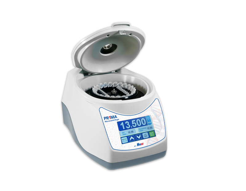
Centrifuges
Spinning: Mini, micro, plate, universal, variable speed, fixed angle, swing-out, ambient, &/or refrigerated, we have a huge range with some of the oldest names in centrifugation.
-

Gloves & PPE
Need it for the lab or cleanroom, we have it: Nitrile, latex, sterile gloves, masks, shields, gowns, coveralls, coats, footies, wipes, & more.
-

PCR / qPCR Reagents
Trusted names at great prices for taq, qPCR master mixes (probe or SYBR substitute), ladders, dNTPs, cDNA, proof reading, RT-PCR, high fidelity, hot start, stains, & more.
-

Conical Tubes
0.5 to 500 mL centrifuge tubes at great values available from MIDSCI. Great names like PR1MA, TPP, Labcon, & more
-

Hoods & PCR Workstations
Crime, chemical, PCR, cell culture, & molecular hoods can be found here.


Why shop MIDSCI?
Founded in 1983, MIDSCI is a leading distributor of lab equipment, scientific supplies and reagents. Our goal is to empower science by delivering innovative and quality products that bring not only value, but also process improvement and optimization.
We understand the importance of your research and maximizing the effectiveness of your funding dollars. Our knowledgeable customer service representatives and product experts are eager to assist with your scientific research questions.
Don’t take our word for it!
"Hello, I am not too sure who to direct this but I wanted to say that your sales rep is fantastic. He is always quick to respond to emails, get me a quote and is just a friendly person. In fact, I have switched from ordering most of my products from Fisher to MIDSCI, all because of his positive and helpful attitude (your products are great too)"
Barbara, St. Louis University
Tariffs Disclaimer:
Due to recent fluctuations in tariffs, pricing is subject to change without notice.
For the most accurate and up-to-date pricing, please contact us directly for a quote.
Web Content Viewer
Actions